2025

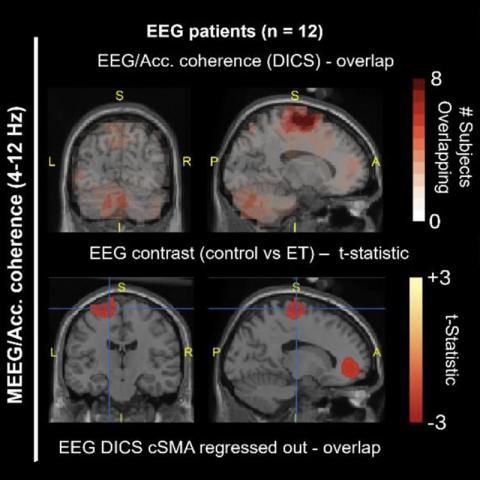
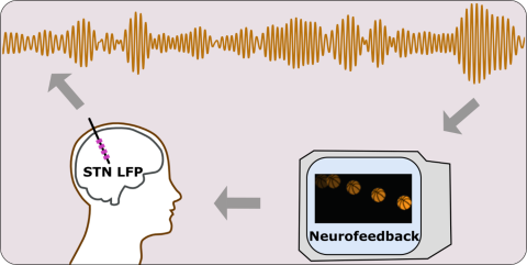
West TO, Steidel K, Flessner T, Calvano A, Spedden ME, Pedrosa D, Barnes G, Cagnan H
10.60964/bndu-k5gj-9892

2024

An image of a person in three states: learning, rest and testing, the figures in 'learning' and 'rest' have headphones on, playing sounds. There are jumbled letters above the head of the 'learning' person, who is reading. The rest person is not reading, and above them there are links forming between nodes, the top row of nodes shows A B C, but not yet with links formed between them. In the final panel, 'Testing', which also shows a light being on, there person has a linked network of nodes, ABC being linked
10.60964/bndu-wv4h-0734
Diagram showing head in profile, with Electrophysiology trace above, as the output from a DBS electrode recording, and neurofeedback on a display shown to the person.
10.60964/bndu-4jde-7j28
A diagram showing PPN DBS electrode recording, power analysis and gait analysis.
10.60964/bndu-w6mx-gv64
a diagram showing the screens that participants see during the trails- in the go, conflict and no-go situations, including what the joystick positions are.
Mandali A, Torrecillos F, Wiest C, Pogosyan A, He S, Tan H, Stagg CJ, Cagnan H
10.5287/ora-qqd05nv46
Image showing the 3 phases of the inference task used. Associative learning phases shows 3 sound icons with arrows pointing to one shape. Conditioning phase shows one shape with an arrow pointing to a pound coin. Inference test phase shows a sound icon with an arrow pointing to a symbol showing a pound coin and wood coin with a question mark. Above the associative learning and conditioning phases there are icons showing musical notes and a coffee cup. The background of the image is two coffee cups on a tabl
Shearer C, Rawson AB, O'Reilly JX, Barron HC
10.60964/bndu-zy02-1973

2023

diagram of a person facing away from the viewer, with representation of deep brain electrodes in their head, with and illustatraion of the task's screen bing displayed to them, and a grasp-strength measuring device in their hand
Herz DM, Groppa S, Brown P
10.5287/ora-9ovjdypbb
diagram explaining the abstract of the paper, see link to paper and the abstract on that page.
Schoenfeld MJ, Thom J, Williams J, Stagg CJ, Zich C
10.5287/ora-jne9aa1z0

2022

diagram of the back of a participant in the study, showing the stimulus display with moving points, and the electrode arrange ment- an electrode in each side of the brain, and two recording sites per electrode, with a stimulating contact in between the two. The participant holds grasping measuring apparatus in each hand.
Herz DM, Groppa S, Brown P
10.5287/bodleian:1R9KzGXxM

2021

image of virtual environment, and images of fMRI and fMRI ouputs being read from brain imaging
Koolschijn RS, Shpektor A, Emir UE, Barron HC
10.5287/bodleian:vmJOOm7KD