Subthalamic and thalamic local field potential recordings from patients with cervical dystonia
Wiest C, Torrecillos F, Morgante F, Pereira EA, Tan H
Description
This data set contains data that were used in Wiest et al., 2022.
The files are in MATLAB .mat format. The dataset is split into three .zip files.
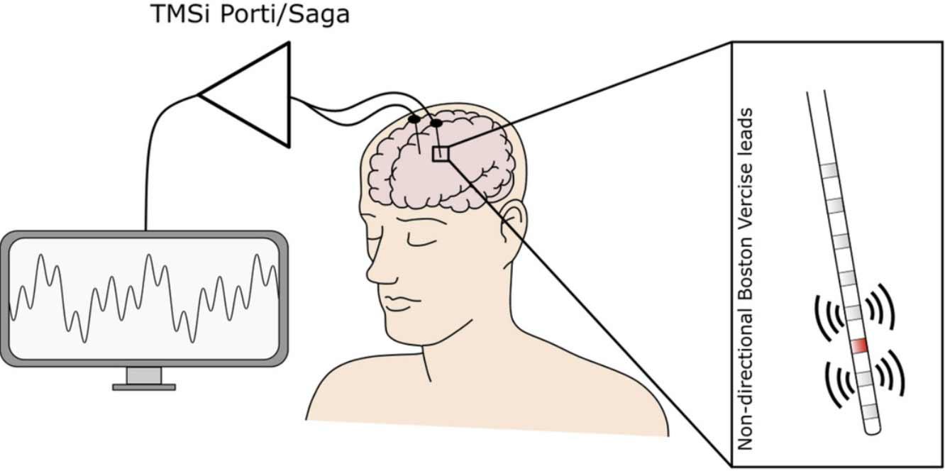
Local field potentials (LFPs) were recorded from 7 patients with cervical dystonia (non-directional Boston Vercise leads with 8 contact levels, placed in the subthalamic nucleus, Zona incerta and ventrolateral thalamus). 5 patients were recorded bilaterally, yielding 12 hemispheres.
All channel labels are saved in SmrData.WvTits. LFPs were recorded from contact levels adjacent to the stimulation contact in bipolar fashion (label e.g. L13 if L2 was stimulated). All other LFPs were recorded in unipolar mode with common mode rejection (label e.g. L4). In addition, selected electroencephalography (EEG) contacts were recorded with common mode rejection (label e.g. C3). In some patients, electromyography (EMG, e.g. left splenius muscles (SplL) or left sternocleidomastoid muscle (SCML)) from dystonic neck muscles and accelerometer data (from the forehead or the vertex) were recorded. The time stamps and intensity of the applied stimulation currents are saved in channel Cur1/Am1.
In this dataset, we progressively increased stimulation intensity in steps of 0.5 mA until either the side effect threshold or 4.5 mA were reached. We tested all middle contacts (levels 2 to 7 if leads are labelled from 1 to 8) in this way.
In Participants 5 to 7, we applied single pulse stimulation (25 pulses) to all 8 contact levels in sequence (file name e.g. P6_Right_Single.mat).
Assistance with this dataset
We welcome researchers wishing to reuse our data to contact the creators of datasets. If you are unfamiliar with analysing the type of data we are sharing, have questions about the acquisition methodology, need additional help understanding a file format, or are interested in collaborating with us, please get in touch via email. Our current members have email addresses on our main site. The corresponding author of an associated publication, or the first or last creator of the dataset are likely to be able to assist, but in case of uncertainty on who to contact, email Ben Micklem, Research Support Manager at the MRC BNDU.
Year Published
2023
Copyright Years
2020-2022
DOI
10.5287/bodleian:QRNQjNpV7
Metadata
Funders
Medical Research Council (MC_UU_00003/2, MR/P012272/1, MR/V00655X/1)
National Institute for Health Research Oxford Biomedical Research Centre (NIHR)
Rosetrees Trust
Publisher
University of Oxford
Downloaded times
Group
Terms
Creative Commons Attribution-ShareAlike 4.0 International (CC BY-SA 4.0)
This is a human-readable summary of (and not a substitute for) the licence.
You are free to:
Share — copy and redistribute the material in any medium or format
Adapt — remix, transform, and build upon the material for any purpose, even commercially.
This licence is acceptable for Free Cultural Works. The licensor cannot revoke these freedoms as long as you follow the license terms. Under the following terms:
Attribution — You must give appropriate credit, provide a link to the license, and indicate if changes were made. You may do so in any reasonable manner, but not in any way that suggests the licensor endorses you or your use.
ShareAlike — If you remix, transform, or build upon the material, you must distribute your contributions under the same licence as the original.
No additional restrictions — You may not apply legal terms or technological measures that legally restrict others from doing anything the licence permits.